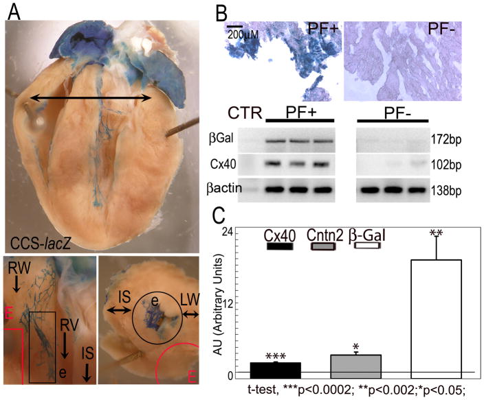
Fig. 1. Identification of novel Purkinje fiber-specific transcripts by comparative microarray analysis.
(A) PF+ and PF− samples were micro dissected distal to the bundle branches (Top, arrow) from the subendocardial and subepicardial regions (Bottom) of CCS-lacZ hearts. (B) Enrichment of PF-specific transcripts in PF+ samples was confirmed by increased expression of the PF markers CCS-lacZ and Cx40 compared to PF− samples, assessed by XGal staining (Top) and/or RT-PCR (Bottom). (C) QRT-PCR showing up-regulation of Cntn2 and the PF-specific markers CCS-lacZ (βGal) and Cx40 in micro dissected PFs. Values are expressed as Arbitrary Units (1AU= RV in PF− sample). PFs, Purkinje fibers; RW, right ventricular wall; LW, left ventricular wall; RV, right ventricle; LV, left ventricle; e, endocardium; E, epicardium. Black outline, subendocardial region; Red outline, subepicardial region; IS, interventricular septum. Size bar: 200μM.

