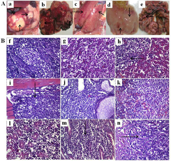
Figure 4.

Clinical and histopathologic observation of diseased chickens in control group infected by MDV LS strain. A. Clinical anatomical change, "a" to "e" were heart, lung, liver, stomachus glandularis, intestinal tract, respectively, and tumors were observed in these tissues, even in the intestinal tract and mesenterium; B. Diagnosis by histopathologic slices (HE 400×). All tissues were fixed in 10% formalin paraffin and embedded, and 5 μm sections were stained with haematoxylin and eosin (HE), for light microscopy, "f" to "n" refer to bursa of fabricius, lung, liver, skeletal muscle, ovary, spleen, kidney, glandular stomach mucosa, cardiac muscle. Arrows show that multimorphology lymphocytes infiltrated patically and formed tumors (h, i, m, n), and other organs were infiltrated by lymphocytes extensively and their organizations were completely destroyed (f, g, j, k, i).
