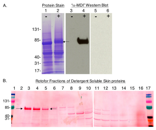
Figure 5.

Detection and fractionation of the major MDI antigen in detergent extracts of exposed skin. (A) Proteins from (-) control or (+) 1% MDI exposed mouse skin, were separated by SDS-PAGE and stained with commassie blue or Western blotted with autologous sera from MDI skin exposed mice (lanes 3 and 4) or control mice (lanes 5 and 6). Arrow highlights major antigenic protein from exposed skin, with apparent shift in migration, indicating change in conformation/charge. (B) The MDI antigen, highlighted by arrows, was separated from other skin proteins by isoelectric focusing. Shown is Ponceau S protein staining of Rotofor® fractions 2-16 after SDS-PAGE and transfer to nitrocellulose membrane. Lanes 1 and 17 contain prestained molecular weight markers.
