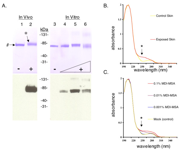
Figure 6.

Purification of antigenically modified albumin from in vivo exposed mouse skin. (A) SDS-PAGE analysis (top) and Western blot with serum IgG from skin exposed mice (bottom) of the major MDI antigen (highlighted with *), purified from skin exposed in vivo to (+) 1% MDI and its corresponding protein purified from (-) control skin (highlighted with #). For comparison, MDI-albumin conjugates prepared in vitro using varying doses of MDI (0.001%, 0.01% and 0.1%, lanes 4 to 6 respectively) are shown to the right of the molecular weght markers. The MDI antigen was not recognized using control sera from vehicle expose mice or irrelevant hyperimmune mouse serum (not shown). (B) Ultraviolet light absorbance spectra of albumin purified from control or 1% MDI exposed skin. (C) For comparison, commercially purified mouse albumin and MDI-mouse serum albumin conjugates prepared in vitro were similarly analyzed. *Note increase in absorbance in the 250 nm range due to MDI's aromatic rings.
