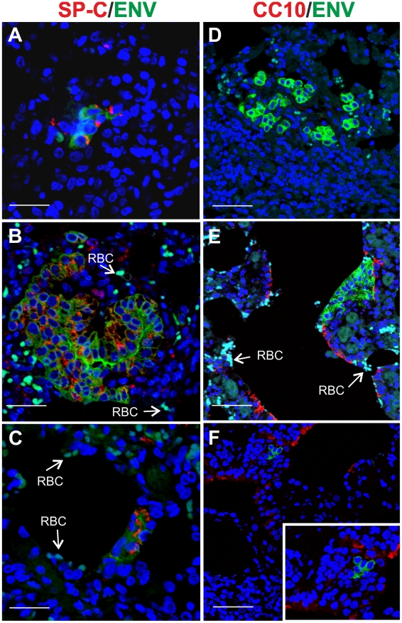
Figure 6. Phenotype of JSRV infected cells in adult sheep with lung injury.
Representative images of lung sections from adult sheep pre-treated with 3MI before experimental JSRV infection. (A–C) Sections were analyzed by confocal microscopy using antibodies towards SP-C (showed in red) and the JSRV Env (showed in green). Nuclei were stained with DAPI and are shown in blue. Arrows indicate autofluorescent red blood cells (RBC). (D–F) Sections analyzed as above using antibodies towards CC10 (showed in red) and the JSRV Env (showed in green). Nuclei were stained with DAPI and are shown in blue. Scale bars, A–B = 47 µm; C = 89 µm; D = 26 µm; E = 33 µm and F = 25 µm.

