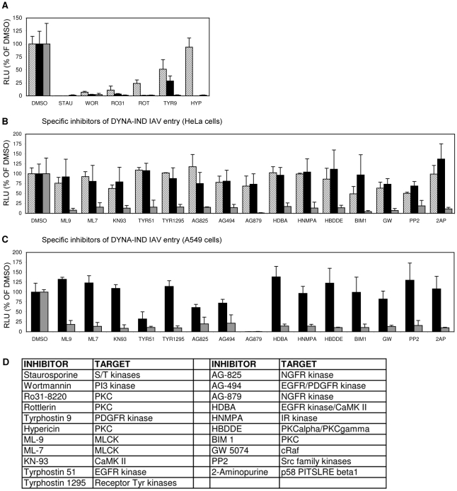
Figure 5. Screening of an eighty-compound protein kinase inhibitor library in an IAV entry assay.
HeLa cells were incubated with kinase inhibitors at a concentration of 10 µM from 1 hr prior to infection (strain WSN; MOI 0.5). Entry (Gluc-entry assay) was performed for 2 hrs in the presence of the inhibitors under DYNA-DEP entry conditions (entry in PBS; black bars) and DYNA-IND entry conditions (entry in PBS supplemented with 10% FCS and 80 µM dynasore; grey bars. In addition, all inhibitors were added 2 hrs p.i. to cells that had not been treated with inhibitors during entry (in presence of 10% FCS) in order to screen for post-entry effects (striped bars). (A) Inhibitors that were inhibitory for both entry routes and mostly also affected post-entry events. (B) Inhibitors that affect DYNA-IND entry but neither displayed effects on DYNA-DEP entry or post-entry effects. (C) All kinase inhibitors were also screened on A549 cells. Black and grey bars correspond to DYNA-DEP and DYNA-IND entry. (D) Targets of inhibition of the different kinase inhibitors are listed. All inhibitors have been tested at a standard concentration of 10 µM. Whereas for some inhibitors higher concentrations may be required for efficient inhibition of their specific target, for other inhibitors this concentration is relatively high and other targets may be inhibited as well. For instance wortmannin is known to inhibit PI4 kinase and MLCK at a 10 µM concentration.

