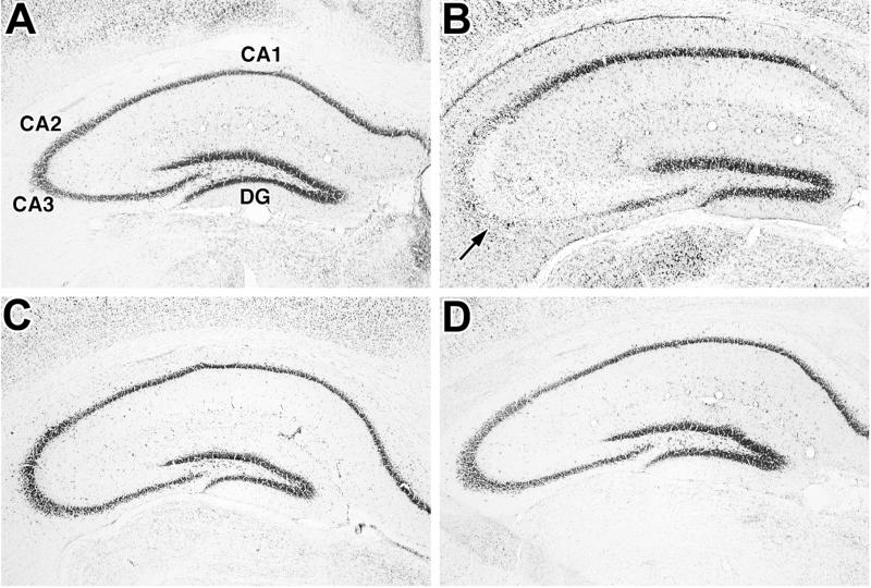
Figure 3.
Comparison of KA-induced damage to pyramidal neurons in the hippocampus of WT (A and B) and SUR1 Tg174 (C and D) mice. Coronal sections of hippocampus were cut from WT and SUR1 Tg mouse brains taken 5 days after administration of saline (A and C) or 25 mg/kg KA (B and D) and stained with cresyl violet. Note the significant loss of pyramidal neurons in the CA2 and CA3 subfields (arrow) of the KA-treated WT mouse (B). No comparable loss of neurons is observed in the KA-treated SUR1 Tg mouse (D). Magnification: ×50.

