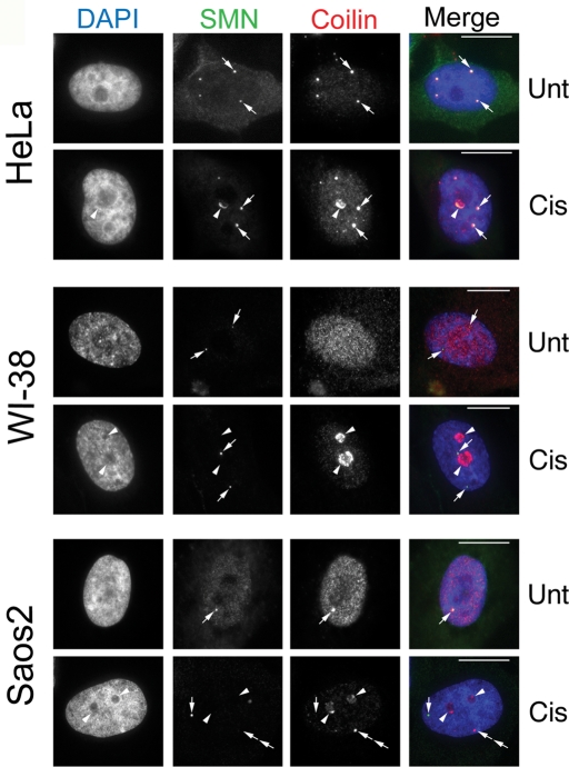
FIGURE 1:
Cisplatin-induced DNA damage triggers nucleolar coilin accumulation. HeLa, WI-38, and Saos2 cells were untreated (Unt) or treated (Cis) for 24 h with cisplatin and immunostained for coilin (red) and SMN (green). Nuclei were stained by 4′,6-diamino-2-phenylindole (DAPI) (blue). For HeLa cells, arrows denote CBs containing both SMN and coilin. Arrowheads mark nucleolar accumulation of coilin and SMN. For WI-38 cells, SMN foci lacking coilin (Gems) are indicated by arrows, and arrowheads mark nucleolar coilin. For Saos2 cells, arrows in the untreated cell denote a CB. Arrows in the cisplatin-treated cells mark a Gem while arrowheads show nucleolar coilin. Double arrows represent an SMN-negative coilin focus. Scale bars = 10 μm.

