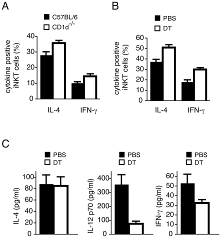
Figure 5. Resident APCs acquire α-GalCer from α-GalCer-loaded BM-DCs in vivo.
(A) BM-DCs derived from C57BL/6j (black bars) or CD1d−/− (white bars) mice were loaded with 200 ng/ml of α-GalCer for 16 h and injected i.v. into C57BL/6j mice. The percentage of splenic iNKT cells expressing intracellular IL-4 or IFN-γ was determined 2 h later by flow cytometry. One representative experiment out of two with three mice per group is depicted, with SEM. (B) CD1d−/− BM-DCs were incubated with α-GalCer as above and injected into lang-DTREGFP mice, which had been injected with either PBS (black bars) or DT (white bars). The percentage of iNKT cells expressing intracellular IL-4 or IFN-γ was determined 2 h later by flow cytometry. One representative experiment out of two with three mice per group is depicted with SEM. (C) Lang-DTREGFP mice were treated with PBS (black bars) or DT (white bars) and then injected i.v. with α-GalCer-loaded BM-DCs from C57BL/6j mice. Serum concentrations of IL-4, IL-12 p70, and IFN-γ were determined 2 h, 5 h, and 10 h later respectively. One representative experiment out of three with 3-5 mice per group is depicted with SEM.

