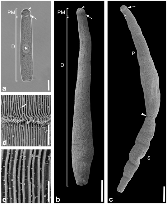
Figure 2. Differential interference contrast (DIC) light micrograph and scanning electron micrographs (SEM) showing the general morphology and surface ultrastructure of the gregarine Heliospora caprellae comb. n.
a. DIC micrograph showing the epimerite (arrowhead), small and rounded protomerite (PM) and the elongated deutomerite (D). The arrow marks the septum between the protomerite and deutomerite. The nucleus (N) is located in the middle of the deutomerite. b. SEM of a single trophozoite with a small protomerite (PM) and a long deutomerite (D) that is wider at the posterior end than at the anterior end. The deutomerite ends in a blunt posterior tip. A slight indentation (arrow) is visible between the protomerite and deutomerite in the area of the septum. Epicytic folds cover the whole trophozoite except for the epimerite (arrowhead) and show an undulating pattern. c. SEM of an association consisting of two trophozoites (or gamonts). The anterior primite (P) has a visible indentation in the area of the septum (arrow) and connects to the anterior end (arrowhead) of the posterior satellite (S). d. Higher magnification SEM of the junction between primite (P) and satellite (S). Some of the epicytic folds (double arrowhead) terminate before they reach the posterior end of the primite. e. High magnification SEM of the epicytic folds. The density of the folds is 5 folds/micron. Scale bars: Fig. 2a, 20 µm; Fig. 2b, 14 µm; Fig. 2c, 11 µm; Fig. 2d, 2 µm; Fig. 2e, 1 µm.

