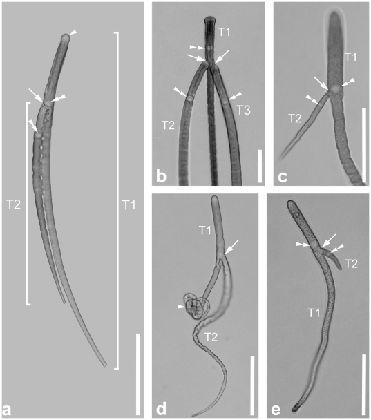
Figure 4. Differential interference contrast (DIC) light micrographs showing the general morphology of the gregarine Thiriotia pugettiae sp. n.
a. DIC micrograph showing an association of two trophozoites. Trophozoite 1 (T1) is longer than trophozoite 2 (T2). The arrowhead marks the slightly broadened anterior tip with the mucron area free of amylopectin. The ellipsoidal nucleus (double arrowheads) is located in the anterior third of the cell in both trophozoites. The attachment site (arrow) of trophozoite 2 is at the level of the nucleus of trophozoite 1. b. Higher magnification view of the anterior end of an association consisting of three trophozoites. The attachment sites (arrows) of trophozoite 2 (T2) and trophozoite 3 (T3) are right behind the nucleus (double arrowhead) of trophozoite 1 (T1). c. Higher magnification view of an association consisting of two smaller trophozoites. The attachment site (arrow) of the much smaller trophozoite 2 (T2) is at the level of the nucleus (double arrowhead) of trophozoite 1 (T1). d. DIC micrograph of two trophozoites. The attachment site of trophozoite 2 (T2) is marked by an arrow. The posterior half of trophozoite 1 (T1) is curled up (arrowhead), a condition that was often recognized when trophozoites were covered in host gut material. e. DIC micrograph of one of the smallest documented trophozoites in an association. The attachment site (arrow) of very small trophozoite 2 (T2) is right behind the nucleus (double arrowhead) of trophozoite 1 (T1). Trophozoite 2 (T2) was only 40 µm long. Scale bars: Fig. 4a, 200 µm; Fig. 4b, 100 µm; Fig. 4c–d, 150 µm; Fig. 4e, 80 µm.

