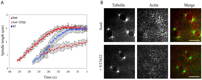
Figure 5. Effect of myosin inhibition on spindle development in Sced embryos.
A) Pole-pole distances in prophase as functions of time (NEB is t = 0 s) show a different degree of pole separation in Sced embryos in the presence and absence of the myosin inhibitor Y27632. The pole-pole separation in Sced embryos is comparable to that in control, but when myosin is inhibited, centrosomes separate less than in control and Sced embryos. B) Images from a time-lapse movie of a Sced embryo in prophase co-injected with FITC-tubulin and rhodamine-actin in the presence or absence of Y27632. Sced embryos have smaller and less organized actin caps which become even less compact when myosin is inhibited. Images are projections of 8–10 confocal planes taken 0.5 microns apart. Red, actin; green, tubulin. Bars, 10 µm.

