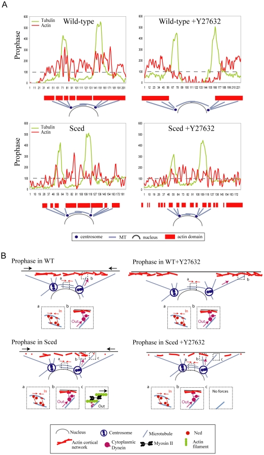
Figure 7. Synergistic actions of actin, myosin and dynein in spindle development.
Hypothesis for each condition (B) is based on the actin distribution as shown in the corresponding line scans (A). The intensity value of 100 has been chosen as the arbitrary cut-off value to discriminate between the ‘soft’ (below 100) and ‘firm’ (above 100) actin patches. In the WT embryo, most of the actin cap is firm, and dynein pulls from the large firm actin cortex (inset b) while kinesin-14 exerts an inward force (inset a). When myosin is inhibited, the area of the firm actin is shifted outward focusing the dynein pulling in the outward direction. In the Sced embryo, many soft actin patches appear, but myosin contracts actin filaments there (inset c) complementing the pulling action of dynein (inset b). When myosin is inhibited in the Sced embryo, dynein pulls from the small infrequent firm patches of actin and does not separate the poles effectively.

