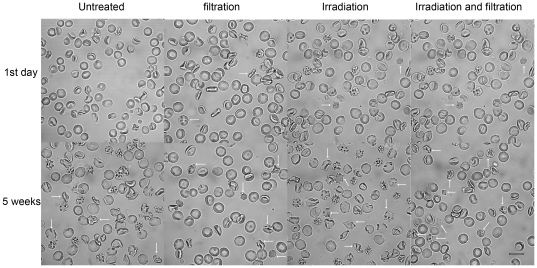
Figure 1. Changes in RBC morphology during storage.
Untreated group, leucocyte filtration group, Irradiation group, Irradiation and filtration group after storaged 1 day and 35 days were observed (white arrow indicated deformation and lesion RBC). Images were taken under Zeiss 510 META confocal laser scanning microscopy (original magnification ×1000, scale bar = 10 µm).

