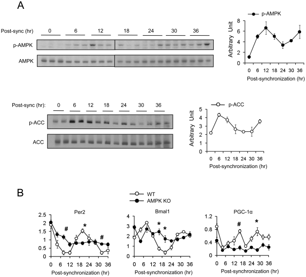
Figure 2. Rhythmic expression of AMPK activity is cell autonomous.
(A) WT mefs were synchronized by forskolin and harvested at the indicated time point. Phosphorylated AMPKα (T172) and phosphorylated-ACC (S79) were assessed by Western blot. Average AMPK and ACC activity (arbitrary unit) were calculated by densitometric quantification of phosphorylated proteins normalized to total proteins. Experiments were repeated at least three times. (B) Expression level (arbitrary units) of mPer2, Bmal1 and PGC-1α in WT and AMPKα1/α2 double knockout (AMPK KO) mefs after synchronization with forskolin. Results are means ± S.E. * P<0.05, # P<0.001between WT and AMPK KO mefs.

