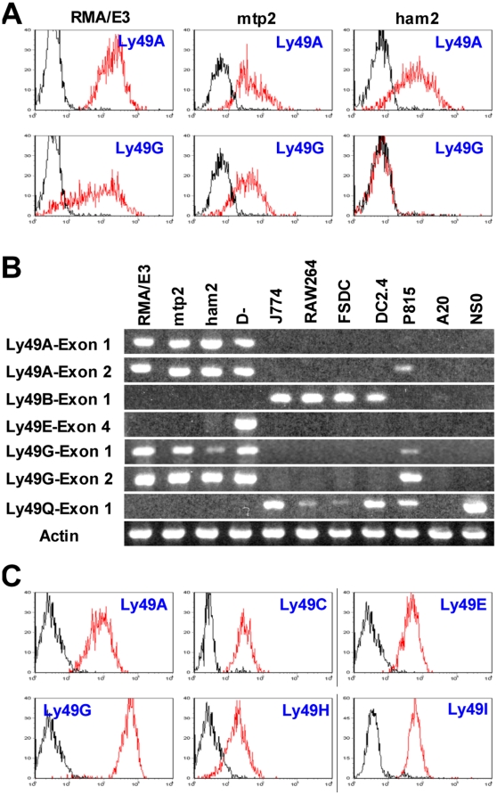
Figure 3. Expression of Ly49 genes in various cells.
A. Different sublines of RMA EL4 cells were stained with JR9 anti-Ly49A and 4D11 anti-Ly49G mAbs (red lines) or medium (black lines) followed by AF647-conjugated secondary antibody. B. RT-PCR analysis of Ly49 transcripts in various cell lines. cDNA prepared from equal numbers of cells was amplified using forward primers located in exon 1, exon 2, or exon 4 together with appropriate reverse primers that in combination gave specific amplification of the relevant Ly49. The identity of the Ly49A exon 1 and Ly49G exon 1 amplimers in RMA/E3 cells was confirmed by cloning and sequencing, as was the unexpected presence of Ly49A, G, and Q transcripts in P815 cells, and of Ly49Q transcripts in NS0 cells. C. D− NK cells were stained with mAbs against the Ly49s shown (red lines) or with medium (black lines).

