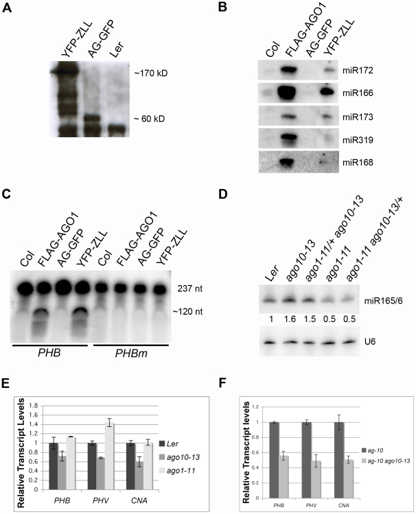
Figure 5. AGO10 is associated with miRNAs in vivo and is catalytically active as a “slicer” in vitro.
(A) Immunoprecipitation (IP) followed by western blotting using anti-GFP antibodies from three genotypes as indicated. The immunoprecipitated YFP-ZLL fusion protein could be detected as a band of the expected size (∼170 kD). AG-GFP (∼60 kD) and Ler served as a positive and a negative control for the IP, respectively. (B) Detection of miRNAs by northern blotting from FLAG-AGO1 and YFP-ZLL IP. Col and AG-GFP served as negative controls for FLAG-AGO1 and YFP-ZLL, respectively. (C) An in vitro “slicer” assay. Both FLAG-AGO1 and YFP-ZLL immune complexes cleaved the PHB wild-type probe, but not the miR165/166-resistant probe. The full-length PHB and PHBm probes were 237 nt. The 5′ and 3′ cleavage fragments were 112 nt and 125 nt, respectively, and were not separated in the gel (represented by the band of approximately 120 nt). (D) Northern blot for miR165/166 in the genotypes as indicated. U6 served as a loading control. The numbers below the gel image indicate the relative abundance of miR165/166. (E) Expression of the HD-Zip genes PHB, PHV and CNA in wild type (Ler), ago10-13, and ago1-11 inflorescences as examined by realtime RT-PCR. (F) Realtime RT-PCR to determine the levels of PHB, PHV, and CNA mRNAs in ag-10 and ag-10 ago10-13 inflorescences.

