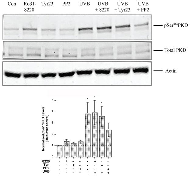
Figure 4. Inhibitors with specificity against Src family tyrosine kinases abrogated UVB-induced PKD activation.
Near-confluent primary mouse keratinocytes were pretreated for 2 hrs with or without the inhibitors Ro 31–8220 (3 μM), tyrphostin 23 (10 μM) or PP2 (10 μM) as indicated and then subjected to sham or UVB irradiation with 30 mJ/cm2. The cells were further incubated for 2 hrs with or without the inhibitors. The cells were lysed and processed for western blotting employing antibodies against phosphoserine916 PKD and total PKD. Actin served as the loading control. Illustrated in the upper panel is a blot representative of 3 separate experiments. Shown in the lower panel are the phosphoserine916 PKD levels from 3 experiments, normalized by total PKD levels and expressed as the means ± SEM; *p<0.05 versus no UVB treatment by a repeated measures ANOVA followed by a Student-Newmann-Keul’s post-hoc test.

