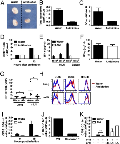
Fig. 4.
Respiratory tract DCs fail to migrate to the draining LN and prime T-cell responses in antibiotic-treated mice. C57BL/6 mice were given antibiotics in drinking water for 4 wk before intranasal infection with 1,000 pfu of PR8-GP33 viruses. (A–C) Three days later, CD11c+ DCs were isolated from the mediastinal LN. (D–F) Naïve p14 tg CD8 T cells (2 × 10⁵ cells per well) were cocultured with different numbers of CD11c+ DCs isolated from mLN of infected animals with (F) or without GP33 peptide (D and E) for 72 h. Splenic DCs from infected animals were used as negative control (E). IFN-γ production (E and F) and the number of CD8 T cells (D) were measured. The number of CD103+ DCs (G) and phenotype of total DC population (H) in the lung and mLN were measured in antibiotic-treated mice without influenza infection. (I and J) Water-fed, antibiotic-treated, and caspase-1–deficient mice were inoculated intranasally with CFSE. Six hours later, mice were infected with 1,000 pfu of PR8 viruses. Eighteen hours after infection, mediastinal LNs were collected. The numbers of CFSE+CD11c+DCs are shown. (K) LPS inoculation (intranasal or intrarectal) restored DC migration to the mLN following intranasal influenza virus infection. Data represent the mean ± SD. Similar results were obtained from three separate experiments.

