Figure 3.
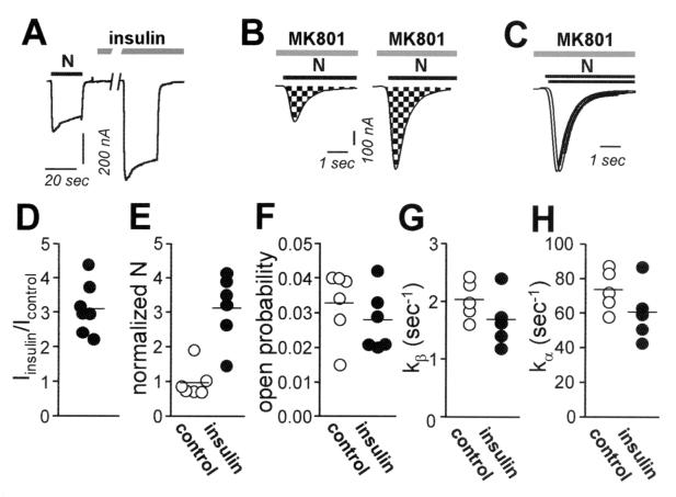
Insulin delivers new functional NMDA channels to the oocyte surface. (A) Whole-cell recordings were obtained from Xenopus oocytes expressing NR1–4a/NR2A receptors in Ca2+-free (Ba2+) Ringer's solution. Insulin (10 min) potentiated NMDA-elicited currents. Insulin, 1 μM; NMDA, 1 mM; glycine, 50 μM. Vh = −60 mV. (B) Insulin increased the number of functional NMDA channels per cell, N. NMDA currents were recorded in the continuous presence of MK-801 (5 μM) from control (Left) and insulin-treated (Right) oocytes; The NMDA-elicited current increased to a peak value, and then decayed exponentially as channels opened and were blocked by MK-801. The cumulative charge transfer, Q, was obtained by integration of the current trace over time (area indicated by checkerboard pattern). The larger integrated current observed for insulin-treated oocytes indicates increased N. (C) Agonist-evoked currents in B were normalized to the peak current to enable analysis of time constants for decay. The time constant of decay of the NMDA current did not differ for insulin vs. control oocytes, indicating no change channel opening rate, k. (D–H) Quantitation of data in A–C. (D) Ratio of NMDA-elicited currents for insulin-treated vs. control oocytes. (E) Channel number per cell, N, normalized to the initial current, I (to correct for variation in expression levels), for control (●) and insulin-treated oocytes (○). (F) Open probability, Po, for control and insulin-treated oocytes (G and H). Opening rate, kβ, and closing rate, kα, did not differ significantly for control vs. insulin-treated oocytes.

