Figure 6.
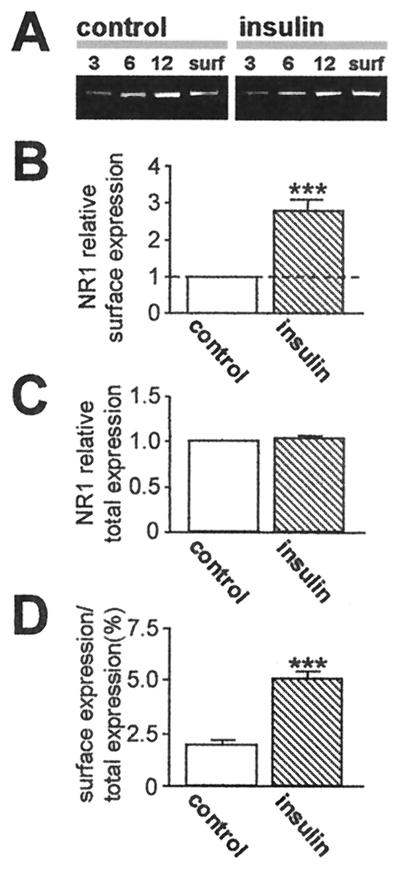
Insulin increases NR1 abundance at the oocyte surface. NR1 surface and total cell expression in control and insulin-treated oocytes, as assessed by Western blot analysis of biotinylated surface protein. (A) Representative Western blot of surface protein from control and insulin-treated oocytes expressing NR1–4a/NR2A receptors probed with anti-NR1 antibody 54.1 (38). Lanes 3, 6, and 12 indicate micrograms of protein in samples of total cell extract before Neutravidin bead extraction loaded on each lane; surface (surf) indicates an aliquot of Neutravidin bead-isolated protein. Insulin increased NR1 abundance in samples of surface protein. (B–D) Quantitative analysis of the effects of insulin on surface expression (B), total cell protein (C), and fractional surface expression (D) of NR1. Mean densities of surface bands were normalized to values for control samples run on the same gels. NR1 subunits expressed at the cell surface increased from 2.0% to 5.3% of total cell NR1; total cell NR1 was unchanged.

