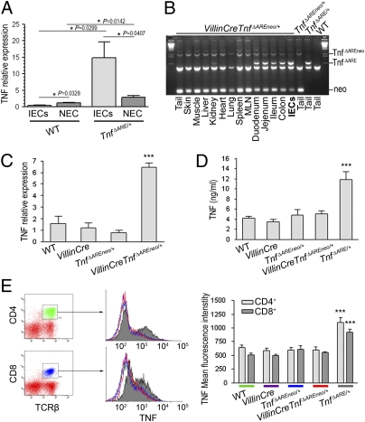
Fig. 1.
IEC are the major early source of TNF in the ileum of TnfΔARE/+ mice. VillinCreTnfΔAREneo/+ mice overproduce TNF specifically by IEC. (A) Quantitative RT-PCR analysis comparing TNF expression in primary ileal IEC and the respective nonepithelial compartment (NEC) in individual mice. Littermate 4-wk-old WT (n = 5) and TnfΔARE/+ (n = 7) mice were used. P values were calculated with two-tailed t test, unpaired for comparison of WT vs.TnfΔARE/+ and paired for comparison of IEC vs. NEC. (B) PCR analysis for detection of the activated TnfΔARE mutation in tissues and primary isolated IEC from VillinCreTnfΔAREneo/+ mice. MLN, mesenteric lymph node. (C) Quantitative RT-PCR analysis for TNF expression in primary IEC isolated from 2-mo-old WT (n = 3), VillinCre (n = 3), TnfΔAREneo/+ (n = 3), and VillinCreTnfΔAREneo/+ (n = 4) littermates. (***P < 0.001; one-way ANOVA.) (D) TNF production determined by ELISA in culture supernatants from LPS-stimulated bone marrow-derived macrophages from 2-mo-old mice (n = 3 per genotype). (***P < 0.001; one-way ANOVA.) (E) TNF expression determined by flow cytometry following intracellular staining for TNF in TCRβ+-gated splenic T lymphocytes from 2-mo-old mice (n = 3–5 per genotype). (***P < 0.001; one-way ANOVA.) In B–E, data shown are representative of two independent experiments. In A–E, mice were analyzed individually. Error bars show SEM.

