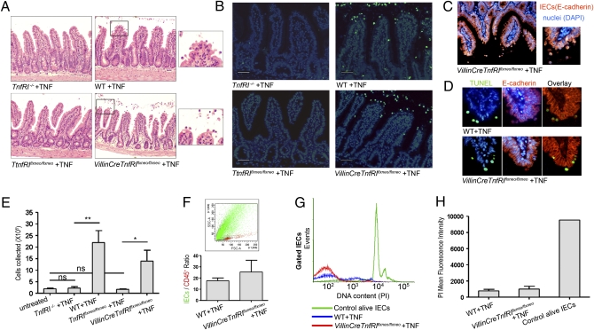
Fig. 4.
Acute exogenous TNF administration leads to IEC apoptosis in a direct manner via epithelial TNFRI. (A–D) Ileal sections from TnfRI−/− (n = 4), WT (n = 5), TnfRIflxneo/flxneo (n = 5), and VillinCreTnfRIflxneo/flxneo (n = 3) mice injected i.v. with 12 μg of murine recombinant TNF. Data represent two independent experiments. (A) Histological examination of H&E-stained sections. (B) TUNEL assay. (Scale bars: 50 μm.) (C) Representative E-cadherin immunostaining. (D) Colocalization of TUNEL and E-cadherin staining in serial paraffin sections. (E–H) Quantitation of TNF-induced IEC apoptosis. Data represent two independent experiments. (E) Number of cells collected from the small intestine of TNF-injected mice. P values (**P < 0.01, *P < 0.05) were calculated with two-tailed t test. Error bars show SEM. (F) Representative FACS analysis for the detection of the markers E-cadherin (epithelial) and CD45 (hemopoietic) on the cells collected. Mean ratio of E-cadherin+/CD45+ cells. Error bars show SEM. (G) DNA content analysis with propidium iodide (PI) staining in gated E-cadherin+/CD45− cells: representative histograms. (H) Quantitation of PI staining data. Error bars show SD.

