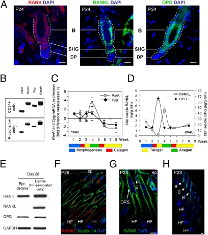
Fig. 3.
Expression of RANK, RANKL, and OPG in HF stem cells. (A) Localization of RANK, RANKL, and OPG in WT telogen bulge (B) and SHG stem cells by immunofluorescence. Nuclei were colored with DAPI. (Scale bars, 20 μm.) Six mice were analyzed for RANK, RANKL, and OPG. (B) RT-PCR of Rank, Rankl, Opg, and Gapdh in FACS-sorted CD34+ bulge and P-cadherin+ SHG cells. (C and D) Measures (mean ± SEM) of Rankl and Opg gene transcription by quantitative RT-PCR in whole skin (C) and of soluble RANKL and OPG by ELISA in skin-organ cultures at the indicated ages (D). n, number of mice for the analysis, a minimum of six mice per time point. (E) RT-PCR of Rank, Rankl, Opg, and Gapdh transcripts in the IFE and the HF-associated dermal cells at P28. (F) Immunofluorescence of RANKL and keratin 14 on a P28 skin section. Nuclei were colored with DAPI. (G) Immunofluorescent labeling for RANK in a P28 skin section with DAPI counterstaining. (H) Immunofluorescent labeling for OPG with DAPI coloration in P28 anagen skin. (Scale bars, 50 μm.) ORS, outer root sheath; B, bulge; ep, IFE.

