Figure 1.
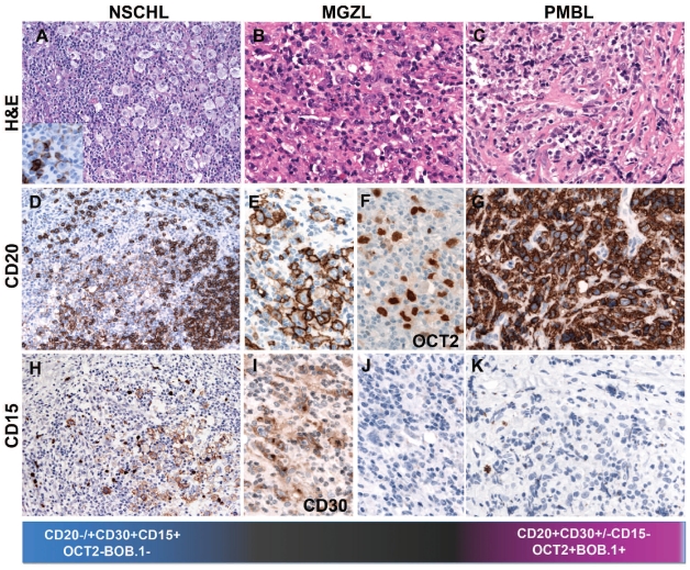
Biological continuum between CHL and PMBL. (A) Nodular sclerosis, classical Hodgkin’s lymphoma with abundant Hodgkin and Reed-Sternberg (RS) cells of lacunar type (H&E, 400x, insert: CD20 immunostain, 400x magnification). The RS cells are weakly and heterogeneously CD20 positive, in contrast to the strong CD20 positivity of the surrounding reactive B cells (immunostaining, 200x). The lacunar cells are CD15 positive (Immunostaining, 200x). (B) Mediastinal gray zone lymphoma with morphological features reminiscent of CHL but an immunophenotype more consistent with PMBL. Note the strong, homogeneous CD20 and OCT2 positivity. The tumor cells are CD15 negative (H&E and immunostaining, 400x). (C) Primary mediastinal B-cell lymphoma composed of an infiltrate of large cells with round or lobulated nuclei and abundant clear cytoplasm. In the background there is a characteristic compartmentalizing sclerosis (H&E, 400x) The tumor cells are CD20 positive and CD15 negative (immunostaining, 400x).

