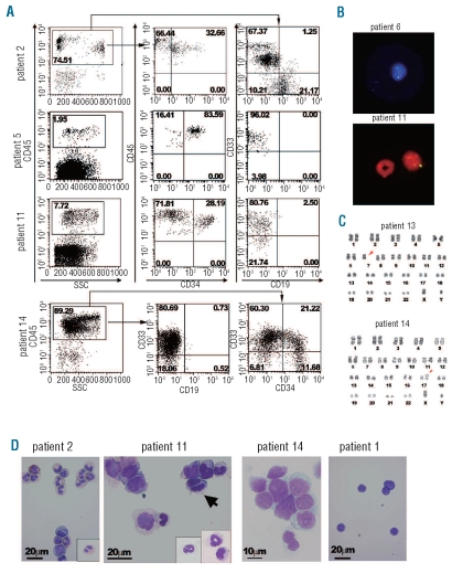
Figure 1.
Engraftment of human MDS-originated hematopoietic cells in the bone marrow (BM) of NOG mice. (A) Representative flow cytometric profiles of BM cells recovered from mice engrafted with patients’ BM cells. The majority of human CD45-expressing cells were positive for a myeloid marker CD33 in patients 5, 11, and 14, while some CD19+ cells were present in BM cells recovered from the mouse engrafted with cells from patient 2. For patient 14, approximately one quarter of CD33+ cells co-expressed CD34. The percentages of cells in the respective regions are shown. (B) FISH detection of a partial deletion of chromosome 7 and monosomy 7. Human cells recovered from the mice engrafted with BM cells from patient 6 and patient 11 were subjected to FISH analysis using D7Z1 (green signal for centromere of chromosome 7) plus D7S486 (red signal for 7q31 region) probes for patient 6 and D7Z1 (yellow signal) probe for patient 11. In a lower panel, a murine granulocyte with a ring-shaped nucleus which did not hybridize with the human probe is located adjacent to the human cell hybridized with D721. All cells analyzed (10 cells for patient 6 and 100 cells for patient 11) demonstrated the same outcome. (C) Chromosomal analysis of cells recovered from the mice transplanted with MDS-originated cells obtained from the BM of patient 13 and patient 14 demonstrated the maintenance of the original abnormal karyotype, namely isochromosome 17 and monosomy 7 (arrows), respectively. Eight cells were analyzed for patient 13 and 20 cells for patient 14. (D) Wright-Giemsa-stained cytospin preparations made of CD45-sorted human cells. In the cytospin samples for patient 2, various stages of myeloid lineage cells and an eosinophil are shown. An insert shows a myelocyte with pseudo-Pelger anomaly. For patient 11, an arrow indicates a bi-nucleated myelocyte. Inserts show differentiated neutrophils. The majority of cells found in a cytospin preparation of BM cells obtained from the mice engrafted with cells from patient 14 demonstrated fine chromatin formation and conspicuous nucleoli. Cytospin samples of a normal cell-engrafted mouse (patient 1) were composed of lymphocytes.

