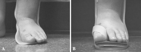
Fig. 3A–B.
(A) A photograph shows a patient in whom the lateral wall of the AFO has been removed. The result is increased pressure across the midfoot and medial malleolus. (B) A photograph shows a patient in whom the lateral wall has been restored with a different AFO. Note improved foot and ankle alignment and improved subtalar alignment.

