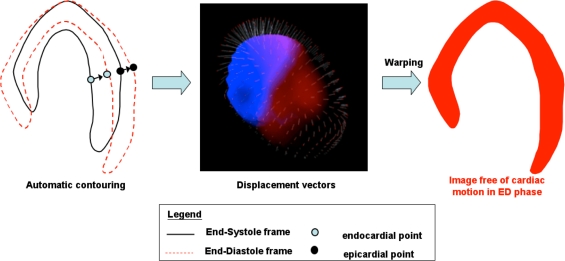
Figure 1.
Diagram illustrating the principle of Cardiac Motion Frozen technique. (Left) Motion tracking of the left ventricle using selected points on endo- and epicardial surfaces. (Middle) Displacement vectors are generated between each cardiac phase. (Right) Starting from those vectors and using a warping technique, all the counts are put back in the end-diastole (ED) phase. The result is an image free of cardiac motion with the noise of a summed static image

