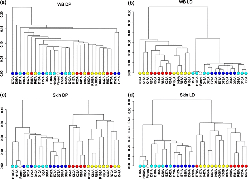
FIGURE 2.

Dendrograms for the electrostatic clustering of barnase alanine-scan mutants based on: (a) WB–DP; (b) WB–LD; (c) Skin–DP; and (d) Skin–LD. Electrostatic potentials were calculated using ionic strengths corresponding to 0 mM ion concentration and εP = 2. Colored circles indicate the free energy change upon mutation for each alanine mutant relative to the parent (white circle). The color code is as follows: red for −500 to −201 kJ/mol, yellow for −200 to −1 kJ/mol, cyan for 1 to 200 kJ/mol, and blue for 201 to 400 kJ/mol
