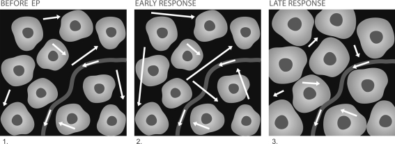
Fig. 1.
An artistic representation of the proposed model of water diffusion in tissue following EP: White arrows illustrate the random movement of water molecules, arrow lengths correspond to the impact-free motion, curved line represents a blood vessel, and light uniform structures on the dark background represent cells, with nuclei shown as dark spots. Before EP: restricted motion; cell membranes are intact. Early response: occurs immediately after onset of the voltage pulse and can last for several minutes after EP, less restricted motion due to increased permeability of the plasma membranes. Late response: occurs minutes/hours after treatment, cell swelling and membrane resealing limit the random motion of the water molecules

