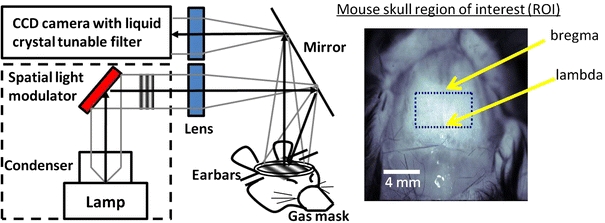
Figure 1.

(Left) Schematic of SFDI imaging of mouse brain through the intact skull. Non-ionizing broadband light is projected onto a spatial light modulator and light intensity patterns of different spatial frequencies are focused onto the mouse’s cranium. The reflectance is captured by a CCD camera with a tunable liquid crystal filter to select for specific wavelengths of light. (Right) Pixels in the region of interest (bregma to lambda and bilaterally to temporalis muscle attachments) are averaged for analyses
