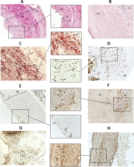
Fig. 2.
Histology of atherosclerotic and preatherosclerotic arteries. H&E and Oil Red staining, together with CD68 and actin IHC were performed with every artery studied in order to identify the artery architecture and to characterize their atherosclerotic lesion degree. Representative images (100× magnification) of a subset of these analyses can be seen in the figure: A, preatherosclerotic radial biopsy, H&E. B, atherosclerotic coronary necropsy, H&E. C, atherosclerotic coronary necropsy, Oil Red. D, atherosclerotic coronary biopsy, CD68. E, preatherosclerotic coronary necropsy, CD68. F, atherosclerotic coronary necropsy, CD68. G, atherosclerotic coronary biopsy, actin. H, preatherosclerotic coronary necropsy, actin. In some images, a subregion has been augmented (200× magnification). I: intima. M: media. A: adventitia. LC: lipid core. FB: fibrous cap. Open-end arrow: VSMCs. Closed-end arrow: macrophages/foam cells.

