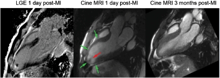
Figure 5.
Viability assessment by late gadolinium enhancement (A) demonstrates the absence of necrosis (lack of bright tissue) in the hypo-akinetic anterior wall [end-systolic image in (B)]. As expected, the follow-up assessment by cardiovascular magnetic resonance demonstrates a major recovery of contractile function in the anterior wall [(C) end-systolic images at follow-up]. The late gadolinium enhancement technique is also sensitive for detection of thrombus, which is attached to a small necrotic (=bright) area in the apex of the left ventricle (A).

