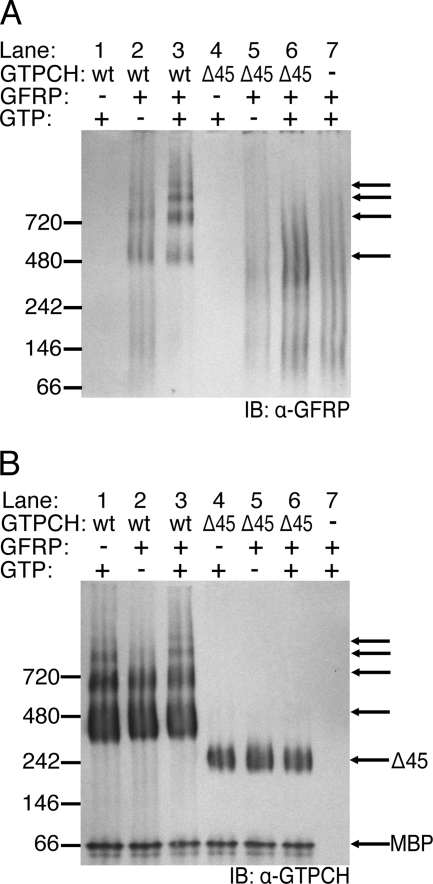
FIGURE 3.
Native complex formation between His6-GFRP and wt-GTPCH or Δ45-GTPCH. wt-GTPCH or Δ45-GTPCH (2.5 μm) were incubated in 100 mm Tris-HCl (pH 7.8) at 37 °C for 1 h. In some reactions, 1 mm GTP and/or 2.5 μm His6-GFRP were also included. A, mixtures containing wt-GTPCH (67.5 ng) or Δ45-GTPCH (55.3 ng) and His6-GFRP (29.5 ng) were resolved on a native PAGE BisTris 4–20% gel before transfer to PVDF membrane and Western blotting (IB) with affinity purified antiserum to GFRP. Black arrows highlight laddering complexes. B, the same PVDF membrane as in A, after stripping and reprobing with antiserum to GTPCH. Unlabeled arrows are as in A, to indicate the position of species identified with anti-GFRP antiserum.

