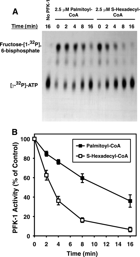
FIGURE 2.
Time course of palmitoyl-CoA- and S-hexadecyl-CoA-mediated inhibition of PFK-1 activity utilizing a direct radiometric assay employing [γ-32P]ATP. Purified rabbit muscle PFK-1 (0.1 μm) was incubated with palmitoyl-CoA (2.5 μm) in 25 mm Tris-HCl, pH 7.5, containing 50 mm NaCl and 1 mm DTT for the indicated times. PFK activity was then assayed in the presence of 1 mm fructose 6-phosphate and 100 μm [γ-32P]ATP (20 μCi/μmol) for 1 min at 25 °C. After termination of the reaction by addition of acetone, the products were resolved by TLC, detected by autoradiography, and quantified by densitometry as described under “Experimental Procedures.” A, representative TLC autoradiograph showing inhibition of fructose [1-32P]6-bisphosphate formation in the absence and presence of palmitoyl-CoA or S-hexadecyl-CoA. B, quantitation of palmitoyl-CoA and S-hexadecyl-CoA mediated inhibition of PFK-1 from three separate experiments (mean ± S.E.) performed as described above.

