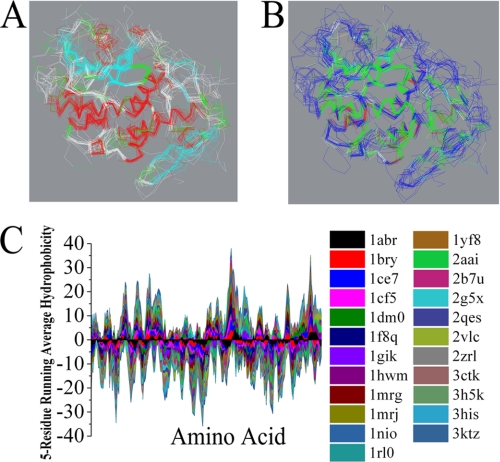
FIGURE 1.
Structure analysis of RIPs. Stereo view of the superimposed backbone atoms for the structures of RIPs. A, secondary structure elements are color coded: helix (red), strand (cyan), and coil (white). B, hydrophobic and hydrophilic residues are colored green and blue, respectively. C, Kyte-Doolittle hydropathy analysis using a window of five amino acids. Hydrophobicity is shown as the vertical axis, with the hydrophobic side of the plot having a positive value. The horizontal axis shows the amino acid residue number along the sequence.

