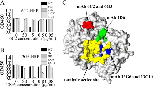
FIGURE 3.
Epitope Identification for RTA mAbs. A and B, microtiter plates were coated with 50 ng/well of RTA. Unconjugated RTA mAbs (5 μg/ml), indicated on the horizontal axis, were added to the coated microtiter wells and incubated for 60 min. The HRP-conjugated Ab indicated at the top of the graph was added, and the mixture was incubated for 60 min. The plates were washed, and colorimetric substrate was added. Antibodies were tested in up to four different experiments, of which this is representative. C, epitope mapping of RTA mAbs. Epitopes defined by mapping using peptide display phage libraries were plotted onto the three-dimensional structure of RTA. Red indicates the epitope of RTA mAbs 6C2 and 6G3, green the epitope of mAb 2D6, yellow the enzyme active site, and blue the residue present in the active site and bound by RTA mAbs 13G6 and 13C10. The graphs are representative of at least three experiments, each of which showed similar results.

