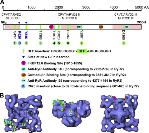
FIGURE 8.
Locations of specific sites and sequences in the three-dimensional structure of RyR2. A, schematic illustration of GFP insertions into various sites in the RyR2 amino acid sequence. The bar represents the ∼5000-amino acid sequence of RyR. Disease-linked RyR1 and RyR2 mutations are largely clustered in three regions (pink shaded areas). In this study, GFP flanked by two glycine-rich spacers was inserted after residues Arg-626 and Tyr-846 (marked by triangle pointed green bands). Other GFP insertions and sites mapped previously are also shown. B, three-dimensional structure of RyR2 with top view (T-tubule face) (left panel), side view (middle panel), and bottom view (sarcoplasmic reticulum face) (right panel). The three-dimensional locations of eight GFP insertions, and GFP inserted after Arg-626 (close to the dantrolene-binding sequence), the FKBP12/12.6-binding site (residues 1815–1855), an anti-RyR antibody (34c) epitope (residues 2722–2769), the CaM-binding site (residues 3614–3643 in RyR2), and an anti-RyR antibody epitope (residues 4377–4494) were shown. The numbers in white indicate subdomains of RyR2. The dashed ellipses indicate regions that potentially contain domain-domain contacts. S437, Ser-437; R626, Arg-626; Y846, Tyr-846; T1366, Thr-1366; T1874, Thr-1874; T2023, Thr-2023; S2367, Ser-2367; T2801, Thr-2801; D4365, Asp-4365.

