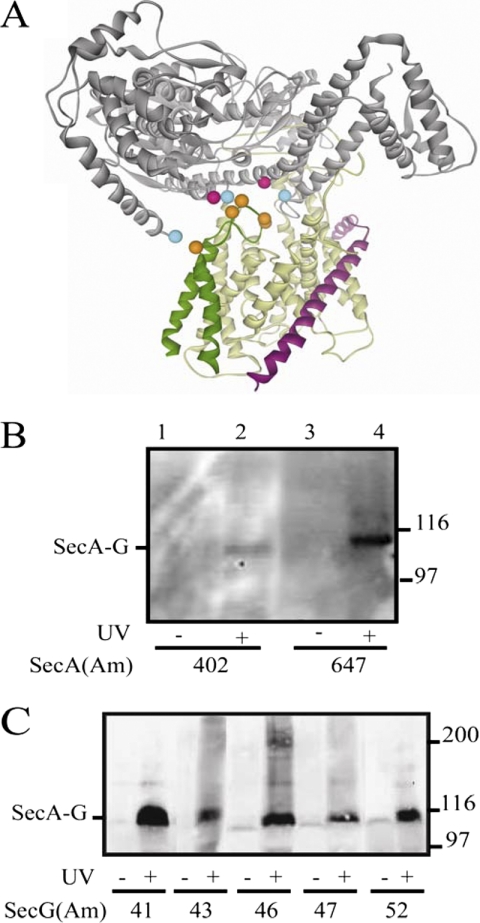
FIGURE 6.
Photocross-linking analysis of secA and secG amber mutant strains. A, SecA is in gray, SecY is in yellow, SecE is in magenta, and SecG is in dark green. The location on the T. maritima SecA·SecYEG crystal structure (38) of residues tested for photocross-linking is depicted as colored balls; SecA residues 402 and 647 that gave positive results are colored in pink, SecA residues 2, 403, and 800 that gave negative results are colored in cyan, and SecG residues 41, 43, 46, 47, and 52 that all gave positive results are colored in orange. B and C, the indicated secA(Am) (B) or secG(Am) (C) mutant strain was grown and subjected to photocross-linking (+) or not (−), and cells were analyzed by Western blotting utilizing c-Myc antibody as described under “Experimental Procedures.” The positions of the SecA·SecG cross-linked complex and molecular weight markers are given.

