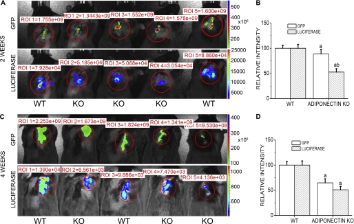
FIGURE 1.
IVIS Imaging of GFP and luciferase signals in murine bone explants model. Kinetic expressions of luciferase and GFP in live animals with double-labeled bone explants were detected at 2 and 4 weeks after explantation. A, GFP and luciferase signal imaging in living animals at 2 weeks post-explantation are shown. B, quantitative analysis of GFP and luciferase signals is shown. C and D, imaging and quantification of GFP and luciferase signals at 4 weeks after explantation are shown. WT, wild type host mice; KO, adiponectin knock out hosts. Results are presented as the mean ± S.E. a, p < 0.05 versus WT; b, p < 0.01 versus WT. ROI, region of interest.

