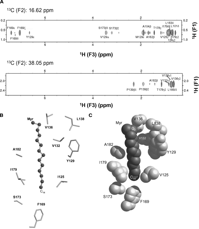
FIGURE 4.
Myristoyl-binding site environment in Ncs1. A, selected slices of 13C(F1)-edited and 13C(F3)-filtered NOESY-HSQC spectra that selectively probe resonances of Ncs1 less than 5 Å away from C14-methyl (top panel) and C2 carbonyl group (bottom panel) of myristate. B, ball-and-stick model of myristoyl group and C-terminal hydrophobic side chain atoms located less than 5 Å away. C, space-filling model of myristate and hydrophobic side chain atoms with same view as in B.

