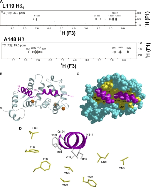
FIGURE 5.
NMR-derived structure of Ca2+-bound Ncs1 bound to Pik(111–159). A, selected slices of 13C(F1)-edited/13C(F3)-filtered NOESY-heteronuclear multiple quantum coherence spectra of 13C-labeled Pik1(111–159) bound to unlabeled Ncs1. B, ribbon diagram of main chain structure of Ca2+-bound Ncs1 bound to Pik(111–159). EF-hands are colored as in Fig. 2. Pik(111–159) is highlighted magenta. C, space-filling representation of Ca2+-bound Ncs1 with same view as in B. D, close-up view of Pik1(111–159) (magenta) with hydrophobic side chains in Ncs1.

