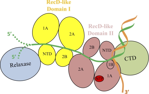
FIGURE 9.
Model of TraI association with a dsDNA-ssDNA junction. The relaxase domain is shown in blue, the RecD-like domain I in yellow, the RecD-like domain II in pink, and the C-terminal domain (CTD) in green. The two strands of dsDNA are colored in orange and dark green. The four subdomains (N-terminal domain (NTD), 1A, 2A, and 2B) of RecD-like domain I and the five subdomains (N-terminal domain, 1A, 2A, 1B, and 2B) of RecD-like domain II are shown and labeled. The known NTP-binding site is highlighted in red. In this model, the 5′ ssDNA overhang binds to the ssDNA binding groove mainly formed by RecD-like domain I and RecD-like domain II. Two possible end structures of the 5′ ssDNA overhang, unattached or covalently attached to the relaxase domain, are presented as dashed lines (47). The 3′ ssDNA tail is not bound by TraI. The dsDNA-ssDNA junction contacts subdomain 1B, or the “pin” domain, of RecD-like domain II.

