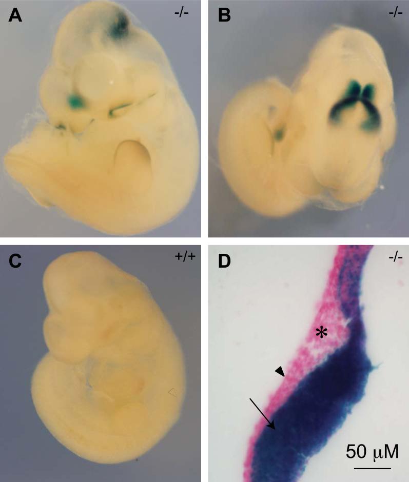
Fig. 3. Af17 promoter activity detected by X-gal staining in E10.5 day embryos.
A-B. An Af17-/- E10.5 embryo shown from side (A) and top aspects (B). X-gal staining is seen in the midbrain-hindbrain junction area (HMJ, or isthmus), margin of the olfactory pit, and the median portion of the most rostral end of the developing forebrain, and tail bud. In addition, weaker staining is seen at the edge of the forelimb bud. C. A WT littermate embryo shown from side aspect with no X-gal staining seen. D. A representative sagittal section of the Af17-/- embryo showing that the expression is limited to the neuroepithelium (arrow). No obvious staining is observed in the ectoderm (arrowhead) and mesoderm (asterisk).

