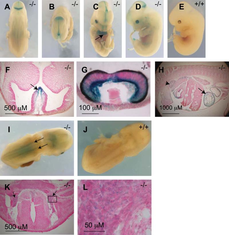
Fig. 4. X-gal staining in E12.5 and E14.5 day Af17-/- embryos.
A-C. At E12.5 day, X-gal staining is seen in the MHJ (A), along the middle of the roof of the midbrain (B) and the median walls of telencephalic vesicles (C). D. Developing optic vesicle begins to show x-gal staining at E12.5 day. E. A wild type embryo showing lack of X-gal staining at E12.5 day. F-G transverse sections confirmed X-gal staining in the median wall of the telencephalic vesicles (F, arrow), eye primordium (G), heart (H, arrow, also C, arrow), and the metanephrone i.e. the developing kidney (H, arrowhead). I. An E14.5 day Af17-/-embryo showing the dorsal root ganglia clearly X-gal stained (arrows). J. A WT E14.5 littermate embryo showing no X-gal staining. K. A transverse section showing the X-gal staining in the dorsal root ganglia (arrows). L. A high magnification image of the boxed area in K.

