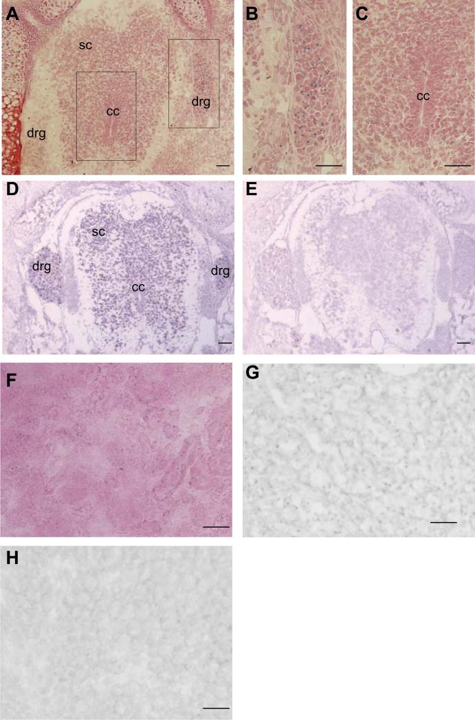
Fig. 8. Af17 mRNA expression pattern detected by in situ hybridization imitates X-gal staining pattern.
A. X-gal staining performed on a transverse section of an E17.5 Af17-/- embryo. An area around the spinal cord and the DRG is shown. Strong staining was observed in the DRG, and relatively weaker staining in the spinal cord. Boxed areas were enlarged and shown in panel B and C. D. In situ hybridization with an Af17-specific anti-sense probe. Signals are identified in the spinal cord and DRG. Very little staining was located in surrounding tissue. E. In situ hybridization with the corresponding sense probe. No clear signal can be identified. F. X-gal staining performed on a kidney section of an E17.5 Af17-/- embryo. Signals can be localized in both cortex and medulla. G and H. In situ hybridization performed on kidney sections with the anti-sense or sense probes. Only the anti-sense probe yielded signals (G), no signal was identified with the sense probe. The sections were counter-stained with brazilin. drg: dorsal root ganglion; sc: spinal cord; cc: central canal. Scale bar: 50 μm.

