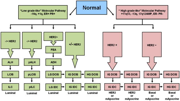
Figure 3.
Divergent evolutionary pathways of breast cancer progression. Genomic and transcriptomic data in combination with morphological and immunohistochemical data stratify the majority of breast cancers into a “low-grade-like” molecular pathway and a “high-grade-like” molecular pathway. The low-grade-like pathway (left hand side) is characterized by recurrent chromosomal loss of 16q, gains of 1q, a low-grade-like gene expression signature, and the expression of estrogen and progesterone receptors (ER+ and PR+). The progression (vertical arrows) along this pathway (green rectangles) culminates with the formation of low and intermediate grade invasive ductal, (LG IDC and IG IDC) and invasive lobular carcinomas including both the classic (ILC) and the pleomorphic variant (pILC). The tumors arising from the low grade pathway are classified as luminal consisting of a continuum of gene expression frequently associated with the absence (luminal A) or presence of HER2 expression (luminal B). The vast majority of ILCs and pILCs and their precursors cluster together within the luminal subtype. The high grade-like gene expression molecular pathway (right hand side) is characterized by recurrent gain of 11q13 (+11q13), loss of 13q (13q−), expression of a high-grade-like gene expression signature, amplification of 17q12 (17q12AMP), and lack of estrogen and progesterone receptors expression (ER− and PR−). The progression along this pathway (red rectangles) includes intermediate and high grade ductal carcinomas that are stratified as HER2, or basal-like, depending on the expression/amplification of HER2. The molecular apocrine subtype, characterized by the lack of ER expression and presence of AR expression, arises from the high grade pathway. The model also depicts intra-pathway tumor grade progression (horizontal arrows). Abbreviations: ALH (atypical lobular hyperplasia), LCIS (lobular carcinoma in situ), pALH (pleomorphic atypical lobular hyperplasia), pLCIS (pleomorphic lobular carcinoma in situ), FEA (flat epithelial atypia), ADH (atypical ductal hyperplasia), Basal (basal-like), mApocrine (molecular Apocrine). +/− HER2 in which the “–”sign is bold indicates that the majority of tumors in the pathway lack HER2 overexpression. (This figure is adapted from [112] ).

