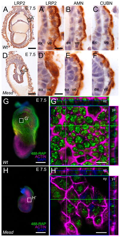
Figure 6. LRP2 requires MESD for apical membrane localization at E 7.5.
Immunohistochemistry indicated LRP2, CUBN, and AMN were apically localized in wild-type VE (A, A′, B, and C). In contrast, LRP2 was distributed diffusely throughout the Mesd-KO VE (D, D′), consistent with ER retention of improperly folded receptor (Hsieh et al.). In contrast, apical localization of CUBN and AMN was not affected by loss of MESD (E, F). Comparison of visceral endoderm endocytosis of 488-RAP in E 7.5 wild-type (G, G′) and Mesd mutant littermates (H, H′) indicated a reduction in functional LRP in Mesd VE. Black boxes in A and D indicate the region magnified in A′, B, C and D′, E, and F. White boxes in G and H indicate the region shown at in G′ and H′. Scale bars in A, D, G, and H indicate 100μm, and in A′, D′, G′, and H′ indicate 10μm. Note that G′ and H′ are shown at the same magnification; however, because orthogonal planes shown in G′ and H′ were chosen to maximize signal from endocytosed 488-RAP, some cells in H′ (or Supplement Figure 2B) appear larger than 488-RAP labeled wild-type due to imaging at a different z-planes. ex – extraembryonic ectoderm, ve – visceral endoderm.

