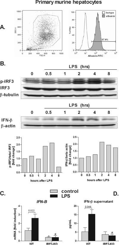
Fig. 4. Parenchymal cells induce Type I IFN in IRF3-dependent manner.
Primary murine hepatocytes were permabilized, stained with anti-albumin antibody, and analyzed by flow cytometry (A). Wild-type primary hepatocytes were stimulated with 100 ng/mL lipopolysaccharide (LPS) for indicated time points and levels of phosphorylated IRF3 (p-IRF3), total IRF3, IFN-β and, beta tubulin and beta actin were analysed using immunoblotting (B). WT and IRF3-deficient (IRF3-KO) primary murine hepatocytes were stimulated LPS, and messenger RNA levels of interferon β (IFN-β) were analyzed by real-time PCR and normalized to 18s (C). IFN-β protein levels in supernatant were analyzed using ELISA (D). Values are shown as mean ± SEM (5 mice per group). Numbers in graphs denote p values; *) p < 0.05 vs. nonstimulated WT hepatocytes; #) p < 0.05 vs. LPS-stimulated WT hepatocytes.

