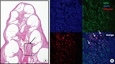
Fig. 4.
Confocal microscope image after immu- nostaining for anti-human nuclei and NF-H. (A) Numerous neurons are observed in the spiral ganglion (black arrow) after 6 weeks of stem cell transplantation (H&E staining, × 40). (B) the expression of anti-human nuclei (red) and NF-H (green) co-localizes with that of DAPI (blue)-positive cells in the spiral ganglion. Grafted stem cells (white arrow) are located in the spiral ganglion (× 200).

